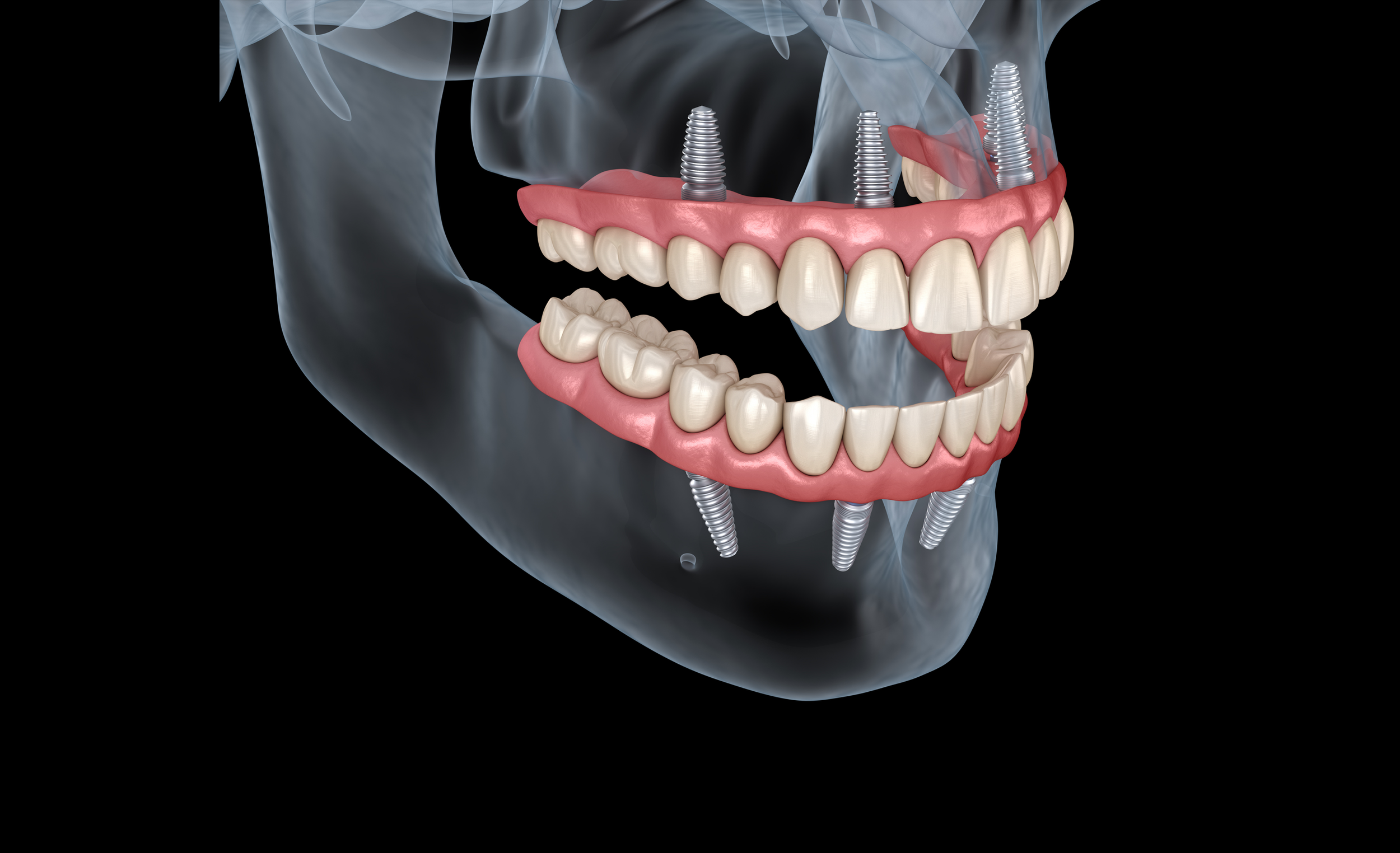
Treatment Planning for Implant Fixed Complete Dentures
Globally complete edentulism varies between regions and countries. More than 240 million patients are partially or completely edentulous (1,2). In the United States, 37 million Americans are edentulous: 26% are between the ages of 65 and 74 (3). Edentulism is more widespread, will remain a challenge, and is a growing public health concern. There is a constant need to restore completely edentulous patients because people live longer. Economically affluent baby boomers account for 20% of the US population, driving future market growth. There will be continued growth of edentulous patients and increased demand for complete dentures in the next 30 years. In 2019 only 314,000 Americans underwent full arch procedures. There is still room for implant fixed complete dentures (IFCD) in our practices. Social media and the internet contribute to the dental education of our patients and make them well-informed about the implant fixed complete denture as a treatment modality. As clinicians, we must follow some guidelines for decision-making to determine if the patient is suitable for such treatment and to ensure the outcome is predictable.
MEDICAL HISTORY
The diagnosis and treatment planning for implant fixed complete dentures begins with a detailed assessment of each patient's medical history and chief concerns. The patient interview is an integral part of the patient's diagnostic workup. Clinicians should ensure that the general health of every patient undergoing elective implant treatment has been thoroughly assessed, given that such treatment includes both surgical and restorative phases.
Check the patient vital signs and review the medication and prescriptions. Suppose significant issues are identified in the medical history. In that case, additional medical assessments should be recommended, some of which may warrant laboratory testing (complete blood count, urinalysis, or sequential multiple analysis of the blood work) before treatment is ever initiated.
Implant dentistry should not be recommended for patients with severe systemic diseases. The primary disease processes that will likely preclude the delivery of dental implant treatment typically are of cardiac or pulmonary origin.
DENTAL HISTORY AND DATA COLLECTION
A patient's dental history, thorough clinical examination, and careful acquisition and evaluation of data that represent the critical findings are needed to arrive at an accurate diagnosis and appropriate treatment plan. Examination of the maxillofacial extra-and intra-oral hard and soft tissues, appropriate preliminary impressions, accurate records required to articulate maxillary and mandibular casts, and routine radiographs and photographs represent vital information that should be carefully collected.
The oral hygiene status should be noted along with the periodontal charting. Determining tissue biotype, bone volume, quality, and the impact of the smile line and the lip line on the esthetic prognosis for the proposed prosthesis are other factors to consider when developing an effective treatment plan for an edentulous patient.
Diagnosis for implant fixed complete dentures for a completely edentulous patient always begins with a thorough clinical extra-and intra-oral examination, a panoramic radiograph, and a Cone Beam Computerized Tomograph (CBCT).
The occlusal vertical dimension (OVD) should be assessed. Occlusal vertical dimension (OVD) changes are observed when teeth are missing. However, evaluation and re-establishment are sometimes tricky, with variations in the dimension. At times, it is difficult to determine the jaw relationships of edentulous individuals. A wax trial denture ("mock setup"), a complete interim denture, or an immediate complete denture should be fabricated to preview the occlusion, vertical dimension, lip support, and esthetics. This setup also provides valuable information regarding the required space needed for IFCDs
Check the vertical space available. Vertical space assessment should begin by measuring the distance between the residual ridge crest and the upper lip or lower lip, depending on the treated arch. It is recommended to measure this dimension with the lip at rest and during a maximal smile. It is important to determine if the edentulous ridge is visible during smiling, as this indicates the prosthesis-mucosal junction will be visible. Such visibility can produce an esthetic liability.
There must be sufficient space available for the desired framework, prosthetic teeth, and denture base material to provide adequate strength, sufficient anchorage for the denture teeth, and be esthetically pleasing. Two millimeters of resin denture base thickness over any metal framework is proposed as the minimum thickness for resin base strength. For a metal-ceramic IFCD, the required vertical space is 15-20 mm, and for All-ceramic IFCD, the required vertical space is 14-16 mm.
DIGITAL TREATMENT PLANNING
Recent advances in technology allowed the laboratory to use computer-aided design/computer-assisted manufacturing technology (CAD/CAM) to fabricate surgical guides that can be either milled or printed. These recent fabrication techniques of surgical guides use DICOM data (Digital Imaging and Communications in Medicine) from the Cone Beam Computed Tomography (CBCT) by importing them into a planning software that allows virtual planning of the implant and the prosthesis. The fabricated guides can be bone supported, mucosa supported, or teeth supported. The guides can be made and the drill holes prepared free-hand, using a milling machine, or using computer-aided design/computer-assisted manufacture (CAD-CAM) technology.
Effective communication is imperative when a team approach that includes multiple practitioners is used. Prior to the treatment plan presentation and informed consent, a consensus should be established between the dental team members, including the laboratory technician. Choosing the proper laboratory is crucial to your success.
*
References:
Peterson PE et al. Community Dent Health 2010.
Moreira Rda S et al. J Appl Oral Sci 2010.
US centers for disease control.
About the author
Dr. Nadim Z. Baba received his DMD degree from the University of Montreal in 1996. He completed a Master’s degree in Restorative Sciences in Prosthodontics from Boston University School of Dentistry in 1999. Dr. Baba serves as a Professor in the Advanced Education Program in Prosthodontics at Loma Linda University School of Dentistry, an Adjunct Professor at the University of Texas Health Science Center School of Dentistry in the Comprehensive Dentistry Department, and maintains a part-time private practice in Glendale, CA. He is currently the President of the American College of Prosthodontists and has received several honors and awards including The David J. Baraban Award from Boston University, the Claude R. Baker Faculty Award for Excellence in Teaching Predoctoral Fixed prosthodontics in 2009 from the AAFP, and the California Dental Association Arthur A. Dugoni Faculty Award in 2010. He published a book entitled “Restoration of Endodontically treated teeth: evidence-based diagnosis and treatment Planning” and has lectured nationally and internationally.


